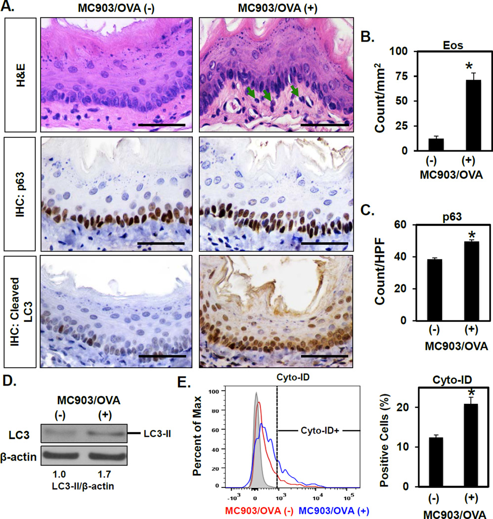
Figure 1. AV content is increased in a food allergy-induced murine model of EoE-like oesophageal inflammation.
BALB/c mice were treated with MC903 and OVA for 18 days (n=3) or untreated as controls (n=4). (A) Oesophageal tissues were evaluated by H&E staining (arrows, eosinophils) and IHC for p63 (basal cells) and cleaved LC3 (autophagy). Scale bars, 50 µm. (B and C) Eos and p63-positive basal cells were determined. Oesophageal epithelial sheets were peeled in (D and E). (D) Immunoblot determined LC3-II (densitometry shown at bottom) with β-actin as a loading control. (E) Flow cytometry determined the Cyto-ID-positive fraction with representative histogram plot (Left) and average (Right) displayed for each condition. *p<0.05 vs. MC903/OVA (−).

