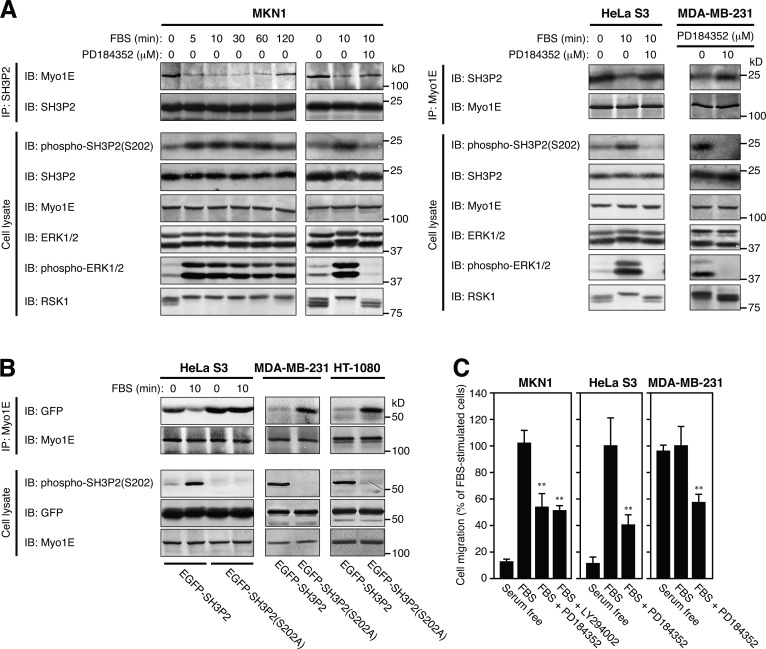
Figure 4.
RSK-mediated phosphorylation of SH3P2 at Ser202 induces the dissociation of Myo1E from SH3P2. (A) MKN1 or HeLa S3 cells were deprived of serum for 12 h, incubated with or without 10 µM PD184352 for 30 min, and then stimulated with 10% FBS for the indicated times. Exponentially growing MDA-MB-231 cells were incubated with or without 10 µM PD184352 for 4 h. Cell lysates were then subjected to immunoprecipitation (IP) with antibodies to SH3P2 or to Myo1E, and the resulting precipitates as well as the whole-cell lysates (10% of the input for immunoprecipitation) were subjected to immunoblot analysis (IB) with antibodies to the indicated proteins. (B) HeLa S3, MDA-MB-231, or HT-1080 cells were transfected for 24 h with vectors for EGFP-SH3P2 or EGFP-SH3P2(S202A), and the transfected HeLa S3 cells were then deprived of serum for 12 h before stimulation with 10% FBS for 10 min. Cell lysates were subjected to immunoprecipitation with antibodies to Myo1E, and the resulting precipitates as well as the whole-cell lysates (10% of the input for immunoprecipitation) were subjected to immunoblot analysis with antibodies to the indicated proteins. (C) The motility of MKN1, HeLa S3, or MDA-MB-231 cells was measured with a transwell migration assay for 6 h in the absence or presence of 10% FBS, 10 µM PD184352, or 10 µM LY294002 as indicated. The numbers of cells that had migrated to reach the lower surface of the filter were 212 ± 20, 63 ± 14, and 177 ± 26 for FBS-stimulated MKN1, HeLa S3, and MDA-MB-231 cells, respectively. Data are representative of at least three separate experiments (A and B) or are means ± SD for three separate experiments (C). **, P < 0.01 versus FBS-stimulated cells.

