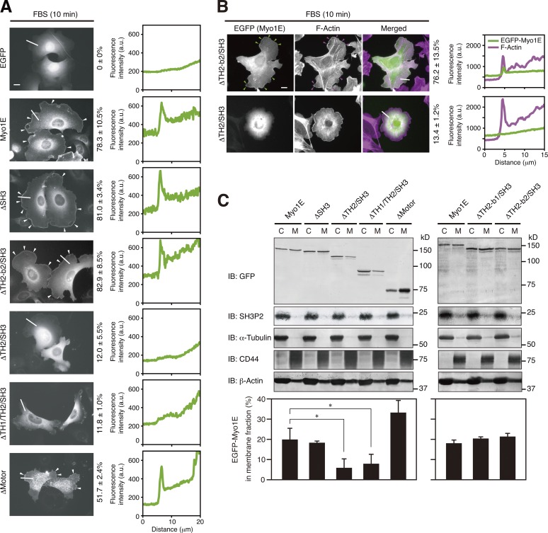
Figure 6.
The TH2 domain mediates the localization of Myo1E to lamellipodial tips. (A and B) MKN1 cells were transfected for 24 h with vectors for the indicated EGFP-tagged Myo1E constructs, deprived of serum for 12 h, and then stimulated with 10% FBS for 10 min. The cells were fixed and then examined for EGFP fluorescence (B, green) with or without staining with phalloidin (B, magenta). White arrowheads (A) indicate Myo1E proteins localized to lamellipodial tips, whereas green and magenta arrowheads (B) indicate colocalization of Myo1E and F-actin, respectively, at lamellipodial tips. Bars, 10 µm. The percentages of cells in which EGFP-Myo1E proteins were localized to lamellipodial tips (A) or colocalized with F-actin (B) are shown at the right of the images as means ± SD for three separate experiments, with n ≥ 80 (A) or 30 (B) cells in each experiment. Fluorescence intensity profiles along the white lines are also shown on the right. Data are representative of at least three separate experiments. (C) MKN1 cells transfected for 24 h with vectors for the indicated EGFP-Myo1E constructs were subjected to subcellular fractionation, and 20% of the cytosolic (C) fraction and 50% of the membrane (M) fraction were subjected to immunoblot analysis (IB) with antibodies to the indicated proteins (top). The proportion of each EGFP-Myo1E construct in the membrane fraction was determined by measurement of immunoblot signals as mean ± SD values for three separate experiments (bottom). *, P < 0.05.

