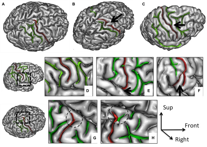
Figure 1.
Morphological features of the central sulcus (CS) with an excellent inter-rater concordance (κ > 0.80) and with values (frequency occurrence) that did not differ from Ono’s post-mortem values. The CS (in red) is represented on a three dimensional (3D) mesh-based reconstruction of the cortex surface. Continuous (A) or interrupted (B, arrow) CS. Connection with the precentral sulcus (PreCS; C, arrow). Inferior end without (D) or with extension to the sylvian fissure (SF; E, arrow). Inferior end “Y” shape (F). Superior end: “T” shape (G) or “Y” shape (H).

