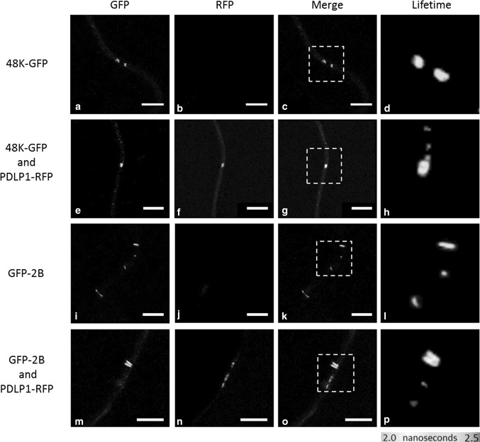
Fig. 2.
Interactions of 48K-GFP and GFP-2B with PDLP-RFP in PDs. Confocal images showing the location and fluorescence lifetime of GFP-labelled CPMV 48K MP, either in the absence (a-d) or presence of PDLP-RFP (e-h). The localisation and lifetime of GFLV 2B MP are also presented in the absence (i-l) and presence of PDLP-RFP (m-p). Reduced fluorescence lifetimes for 48K-GFP in the presence of PDLP-RFP can be seen in h (compare lifetime to d), and GFP-2B in p (compare lifetime to l). Lifetime image panels (right column) display a pseudo-coloured image representing the GFP lifetime, as indicated by the colour scale below the column. White dashed boxes indicate spots portrayed in lifetime image. Scale bar = 5 µm

