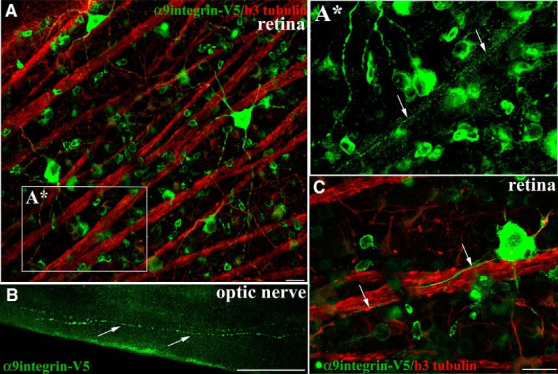
Figure 3.
α9integrin-V5 expressed in adult RGCs is transported into optic nerve axons. Confocal images of flat mount retina show RGCs immunolabeled with anti-V5 (green) and colabeled with anti-β3 tubulin (red) 3 weeks after intravitreal injection of AAV2-CAG-α9integrin-V5 (A, C). A*, A high magnification image (from A) of integrin-containing axons in a fascicle (arrows) travelling toward the optic nerve. Epifluorescent images in B of optic nerve indicate V5-labeled α9integrin within axon fibers of the optic nerve 3 weeks following AAV injection. Arrows in C indicate V5-labeled axons following along the course of β3 tubulin axons. Scale bars: A, C, 20 μm; B, 50 μm.

