Figure 1.

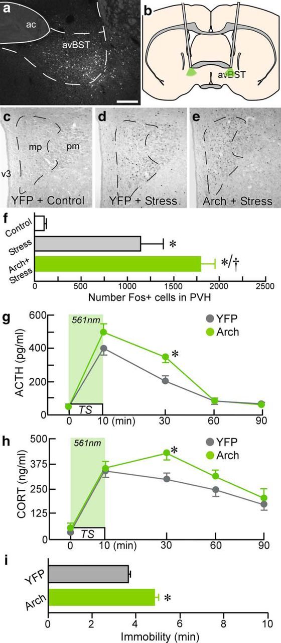
Endocrine and behavioral consequences of avBST somata photoinhibition. a–e, Dark-field image (left) of a coronal section showing YFP-fluorescent neuron soma after AAV microinjection into avBST, with a schematic coronal section (b) illustrating fiber optic placement for the bilateral photoinhibition of neuronal somata therein. Example photomicrographs showing Fos immunoperoxidase labeling in PVH from unstressed YFP control (c), as well as YFP (d) and Arch (e) rats subjected to TS, illustrating a marked increase in Fos immunoreactivity after avBST somata photoinhibition. Scale bar, 200 μm (a, c–e). f, Quantification of Fos-immunoreactive nuclei reveals a significant induction as a result of stress exposure (*p < 0.05) and further enhancement associated with photoinhibition of avBST neuron axons (†p < 0.05). n = 3 YFP + control, n = 7 YFP + stress, n = 6 Arch + stress. g, Analysis of ACTH levels before and after 10 min TS coincident with 561 nm illumination (light green shaded area) of avBST somata. h, Plasma levels of CORT were significantly elevated in Arch animals 30 min after the onset of stress versus YFP controls (*p < 0.05). Arch stimulation resulted in a prolonged elevation of plasma titers of ACTH and CORT (at 30 min) after stress onset compared with YFP control animals (*p < 0.05). i, Photoinhibition of avBST somata was associated with a significant increase in immobility behavior during TS. n = 6 YFP, n = 6 Arch (f–i).
