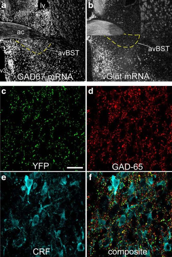
Figure 6.

Phenotypic characterization of avBST neurons and terminal fields in PVH. a, b, Dark-field photomicrograph showing in situ hybridization of GAD67 (a) and VGLUT (b) mRNA in avBST and its vicinity. c–f, Confocal laser-scanning microscopic image displaying the distribution of YFP-fluorescent terminals (green) that originated from avBST (c) and corresponding images in the same z-section showing GAD-65 (d, red) and CRF (e, cyan) immunoreactivity (f, composite). Scale bar, 400 μm (a, b); 20 μm (c–f).
