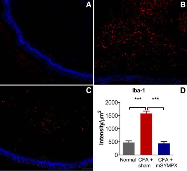
Figure 10.
mSYMPX reduces macrophage staining in the CFA-inflamed paw. Paw skin sections were obtained from normal animals or 4 d after CFA injection (CFA), which was performed 14 d after mSYMPX or sham mSYMPX surgery. Red staining represents macrophage marker Iba-1. Blue represents DAPI nuclear stain. A, Example of Iba-1 staining in normal paw skin. B, Example of CFA-inflamed paw with prior sham mSYMPX. C, Example of CFA-inflamed paw with prior mSYMPX. D, Summary data of normalized Iba-1 intensity. ***p < 0.001, significant difference between indicated groups (ANOVA with Tukey's post hoc test). The normal and LID + mSYMPX groups did not differ significantly from each other. N = 4 male rats per group, 17–21 sections per animal. Scale bars, 100 μm.

