
Fig. 3. Deletion of the repeat region of REX1 decreases the number of Maurer’s cleft puncta.
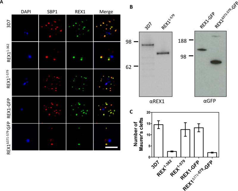
A. Immunofluorescence microscopy of acetone-fixed infected RBCs probed with anti-SBP1 (red) and anti-REX1 (green). Nuclei are stained with DAPI (blue). Scale bar = 3 μm. B. Western blots confirming expression of REX1 in wildtype 3D7 and truncated REX11–579 (probed with anti-REX1), and REX1-GFP and REX1(Δ371–579)-GFP chimeras (probed with anti-GFP). C. Quantitation of numbers of Maurer’s clefts produced by 3D7 and different REX1 transfectants in singly nucleated infected RBCs. Error bars = SD.