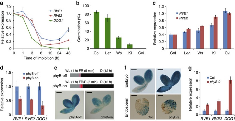
Figure 3. Expression pattern of RVE1 and RVE2.
(a) Relative expression of RVE1, RVE2 and DOG1 during imbibition. Freshly harvested Col seeds were imbibed in 0.6% agar plates for the indicated time. (b) Germination frequency of freshly harvested seeds from Col, Landsberg erecta (Ler), Wassilewskija (Ws), Köln (Kl) and Cape Verde Islands (Cvi) ecotypes. Seeds were grown under white light conditions for 4 days. (c) Relative expression levels of RVE1 and RVE2 in different ecotypes. Total RNA was isolated from seeds imbibed under light for 12 h. (d) Quantitative reverse transcriptase–PCR of RVE1 and RVE2 in post-harvest Col seeds grown under phyB-off and phyB-on conditions. (e) GUS staining of RVE1p:GUS transgenic seeds after phyB-off and phyB-on treatments as indicated in the top panels. D, dark; FR, far-red light; R, red light; WL, white light. (f) GUS staining of freshly harvested Col and phyB seeds harbouring RVE1p:GUS. The seeds were incubated in darkness for 12 h. Scale bars, 200 μm (e,f). (g) Relative expression of RVE1, RVE2 and DOG1 in Col and the phyB-9 mutant. Freshly harvested seeds were imbibed for 36 h in darkness. For a–d and g, mean±s.d., n=3.

