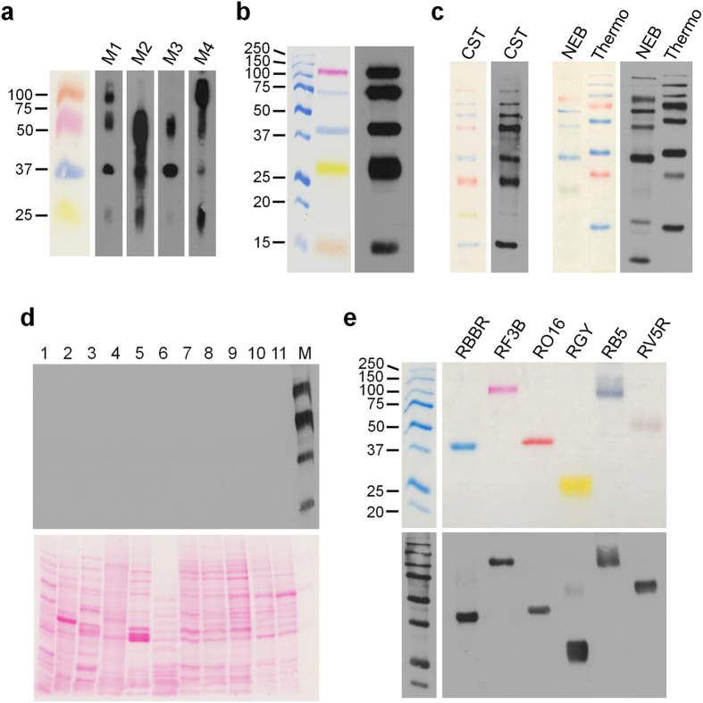
Figure 2. Anti-RAINBOW antibody detects multi-colour Remazol prestained molecular weight markers.
(a) Presence of antibodies against four Remazol dyes in mouse sera. Immune sera of mice (3rd bleed) immunized with different prestained proteins were tested with a Miniblotter 28 channel unit against Remazol dye stained phosphorylase b (orange), γ-globulin heavy chain (red), ADH (blue), and γ-globulin light chain (yellow). M1, M2, M3, M4: mouse 1, 2, 3, 4. 20× more dye stained proteins were loaded on the colorimetric marker lane than on the lanes used for Western blot analysis with the mouse sera. (b) The anti-RAINBOW monoclonal antibody shows equal detection of Remazol-dye stained proteins: phosphorylase b (red), BSA and ADH (blue), chymotrypsin (yellow) and lysozyme (orange). 10× more dye stained proteins were loaded on the colorimetric lane shown in the left panel than on the lane incubated with anti-RAINBOW antibody. (c) anti-RAINBOW monoclonal antibody detects all marker bands in a variety of commercial multicolor protein marker mixtures. CST: Cell Signaling Technology, Color-coded Prestained Protein Marker, High Range (12949P); NEB: New England Biolabs, ColorPlus Prestained Protein Marker (P7709); Thermo: Thermo Scientific: PageRuler Plus Prestained Protein Ladder (26619). 5× more of each protein marker mixture was loaded on the colorimetric lanes with the visible marker bands than on the lanes incubated with anti-RAINBOW antibody. (d) anti-RAINBOW monoclonal antibody does not cross-react with unstained cellular proteins. 1: E. coli; 2: A. thaliana; 3: S. cerevisiae; 4: C. elegans; 5: D. melanogaster; 6: Chicken follicle; 7: CHO, Chinese hamster ovary; 8: Rat1, rat fibroblasts; 9: N2a, mouse neuroblastoma; 10: CV1, Green monkey epithelial; 11: Hela, human epithelial; M: homemade prestained rainbow marker (RF3B-phosphorylase b, RBBR-BSA, RO16-ADH, RGY-γ-globulin light chain). (e) anti-RAINBOW monoclonal antibody cross-reacts with Remazol dyes not used for immunization. Lane 1: RBBR-ADH (blue), lane 2: RF3B-phosphorylase b (red), lane 3: RO16-ADH (orange), lane 4: RGY-γ-globulin light chain (yellow), lane 5: RB5-phosphorylase b (black), lane 6: RV5R-γ-globulin heavy chain (violet). 5× more Bio-Rad marker proteins and 10× more of the Remazol stained proteins were loaded on the upper blot (colorimetric image) than on the lower blot incubated with anti-RAINBOW antibody.

