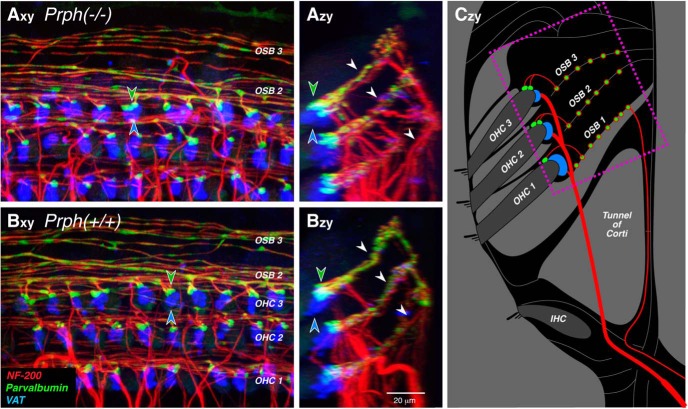
Figure 3.
Immunostaining for parvalbumin (which stains type II terminals and outer spiral fibers green) and vesicular acetylcholine transporter (which stains MOC terminals blue) suggests that both afferent and efferent innervations are normal in Prph−/− ears. A, B, Maximum projections of confocal z-stacks from the 16 kHz region of a Prph−/− ear (A) and a Prph+/+ ear (B), shown in the acquisition plane (x–y) and in the orthogonal plane (z–y) showing a cross-sectional view, as schematized in C. The dotted box in C shows the approximate region imaged in A and B. Green-filled and blue-filled arrowheads in A and B highlight the spatially offset clusters of type II and olivocochlear terminals, respectively, underneath the third-row OHCs. White arrowheads in Azy and Bzy point to the three outer spiral bundles (OSBs) running between the Deiter’s cells. Scale bar: Bzy (for all panels), 20 μm.

