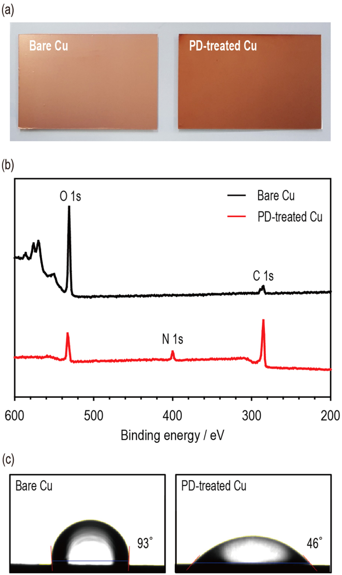
Figure 1. Preparation of the polydopamine-treated Cu current collector.

(a) OM images of the bare Cu (left) and the polydopamine-treated Cu (right). (b) XPS spectra of the bare Cu (top) and polydopamine-treated Cu (bottom). (c) Contact angle images of the bare Cu (left) and the polydopamine-treated Cu (right).
