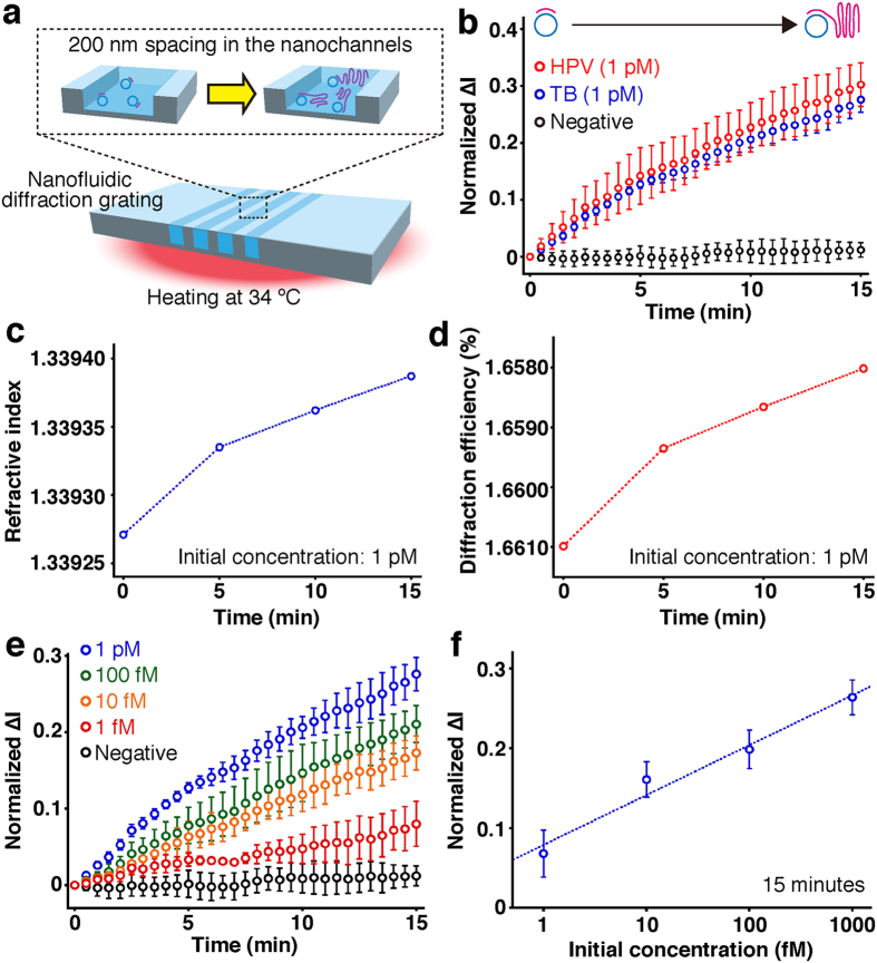
Figure 3. Label-free detection of real-time DNA amplification in the nanofluidic diffraction grating.
(a) A schematic illustration showing real-time DNA amplification in the device at 34 °C. (b) Time-course monitoring of normalized ∆I of the diffracted light during real-time DNA amplification with different target sequences. Target sequences of amplified DNA molecules were human papillomavirus (HPV, red circles, initial concentration: 1 pM) sequence and tubercle bacilli (TB, blue circles, initial concentration: 1 pM) sequence. Negative control data (black circles) were obtained with no target sequence. Error bars show the standard deviation for a series of measurements (N = 3). (c) Refractive index versus amplification time. The initial concentration of TB sequence was 1 pM. (d) Diffraction efficiency derived from RCWA versus amplification time. The initial concentration of TB sequence was 1 pM. (e) Time-course monitoring of normalized ∆I of the diffracted light during real-time DNA amplification for TB sequence with different initial concentrations. Negative control data (black circles) were obtained with no target sequence. Error bars show the standard deviation for a series of measurements (N = 3). (f) Normalized ∆I plot derived from signal changes by DNA amplification for 15 min in the nanochannels, with respect to the initial concentration of TB sequence. Error bars show the standard deviation for a series of measurements (N = 3).

