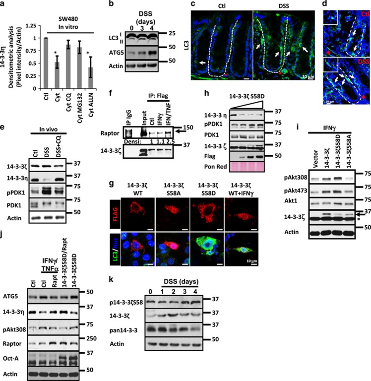
Figure 4.
Autophagy triggered by 14-3-3ζ induces 14-3-3η degradation and Akt activation during inflammation. (a) 14-3-3η protein levels were analyzed in cell lysates of SW480 cells treated with IFNγ/TNFα (Cytokines) in the presence or absence of proteasome (MG132), lysosome (CQ) and Calpain (ALLN) inhibitors. Densitometric analysis is shown in graph. n=3. *P<0.05; **P<0.001. (b) LC3 I-II and ATG5 were analyzed in mucosal cell lysates of C57BL/6J mice exposed to DSS for 1–4 days. Actin was used as a loading control. (c) LC3 (green) and (d) p62 (green) localization was analyzed by immunofluorescence in cryosections obtained from colonic mucosa of control mouse and mice exposed to DSS. C57BL/6J received drinking water or drinking water with DSS (3%) during 4 days. Positive epithelial cells are marked with white arrows. Lamina propria cells that are positive are marked with red star. Crypt axis is marked by white dotted lines. Nuclei are blue. Bar=10 μm. (e) 14-3-3ζ, 14-3-3η, pPDK1 and PDK1 were analyzed in cell lysates of C57BL/6J mice treated for 4 days with DSS (3%). Mice were injected daily with vehicle alone or with Chloroquine (CQ; 40 mg/kg). Actin was used as a loading control. n=3. (f) Association of raptor with 14-3-3ζ was analyzed by co-immunoprecipitation assays. Overexpressed 14-3-3ζ-Flag and control IgG were immunoprecipitated from fresh lysates obtained from SW480 control cells or SW480 cells treated with IFNγ or IFNγ/TNFα for 6 h. Immunoprecipitates were blotted for raptor and 14-3-3ζ. Densitometric values for raptor are shown. (g) Autophagosome formation was analyzed in SW480 cells transfected with 14-3-3ζ WT, 14-3-3ζ S58D, 14-3-3ζ S58A and 14-3-3ζ WT/IFNγ. 14-3-3 proteins are Flag tagged (red). LC3 is marked in green. Nuclei are blue. Bar=10 μm. (h) 14-3-3η, pPDK1, PDK1, 14-3-3ζ and Flag were detected in cell lysates of SW480 cells expressing increasing concentrations of 14-3-3ζ S58D. Ponceau red was used as a loading control. (i) pAkt308, Akt1 and 14-3-3ζ were detected in cell lysates of SW480 cells transfected with 200 ng of 14-3-3ζ WT, 14-3-3ζ S58D or 14-3-3ζ S58A. Cells were treated with IFNγ for 18 h. Actin was used as a loading control. Arrow=overexpressed 14-3-3ζ. Star=endogenous 14-3-3ζ. (j) ATG5, 14-3-3η, pAkt308, raptor and Flag were detected in cell lysates of SW480 cells expressing 14-3-3ζ or 14-3-3ζ/raptorS792A. Twelve hours post transfection, cells were treated with IFNγ/TNFα for 24 h. Actin was used as a loading control. (k) p14-3-3ζS58, 14-3-3ζ and pan14-3-3 were analyzed in mucosal cell lysates of C57BL/6J mice treated with DSS for 3 days. Actin was used as a loading control

