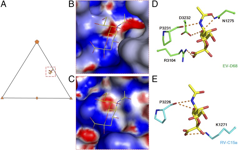
Fig. 2.
A potential binding site for glycans on the RV-C receptor. (A) A triangle indicates an icosahedral asymmetric unit. A red rectangle (dashed line) outlines the limit of the sialic acid binding site shown in B and C. Surface electrostatic potential of EV-D68 (PDB ID code 5BNO) (B) and RV-C15a (C) is represented with a scale of −8kT/e (red) to 8kT/e (blue). (D and E) The sialic acid (yellow) interacts with surrounding residues on EV-D68 (green) and as anticipated on RV-C15a (cyan). Red dashed lines indicate (potential) polar interactions. Oxygen and nitrogen atoms are colored red and blue, respectively.

