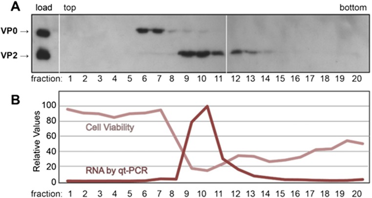
Fig. S2.
Characterization of two forms of RV-C15a particles. A sample of RV-C15a was sedimented through a sucrose gradient. Fractions (1 mL) were collected (from the top) and then probed for VP2/VP0 content by Western blot analyses (A) using mouse anti-RV-C15-VP2. These fractions were also tested for infectivity according to cytopathic effect and for RNA content by qRT-PCR (B).

