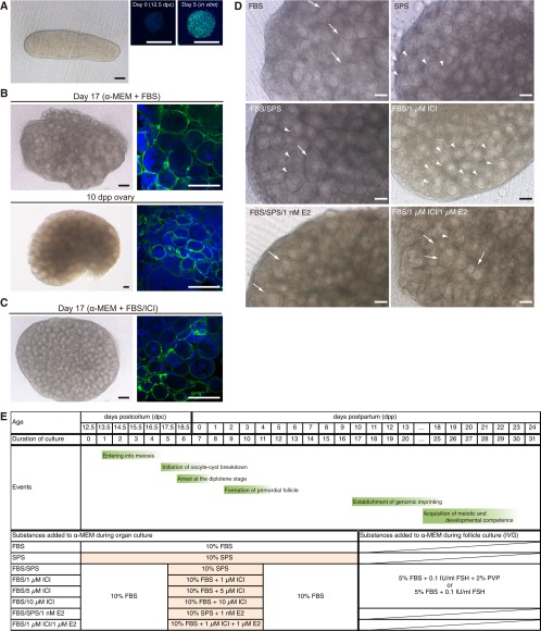
Fig. S1.
Refinement of culture condition for follicular assembly. (A) Gonad derived from a mouse embryo at 12.5 dpc (Left). (Black scale bar, 100 μm; white scale bars, 20 μm.) Immunostaining for SCP3 (green) in female germ cells derived from an ovary on day 0 (Center) and day 5 of culture without the mesonephros (Right). Nuclei were stained with 4′,6-diamidino-2-pheylindole (DAPI; blue). (B) Ovary cultured for 17 d (α-MEM + FBS) and the ovary at 10 dpp (control). Shown are bright-field images (Left) and laminin immunostaining (green) and DAPI staining (blue) of the ovary (Right). (Black scale bars, 100 μm; white scale bars, 50 μm.) (C) Ovary cultured for 17 d (α-MEM + FBS/10 μM ICI). (Black scale bars, 100 μm; white scale bars, 50 μm.) (D) Ovary cultured in the medium indicated. The parts where the border of follicles was not clear and multioocytes were contained in a follicle were indicated by white arrows. The parts where each follicle was clearly visible with a single oocyte were indicated by white arrowheads. Note that the addition of ICI drastically improved the formation of follicles with single oocytes. In contrast, the addition of estradiol disturbed such follicular formation. (Scale bars, 100 μm.) (E) Oogenetic events and examined conditions for the culture. α-MEM was used as the basal medium. The effects of supplementation of FBS, SPS, estradiol (E2), ICI, and PVP were examined. On day 17 of culture, secondary follicles were isolated from the in vitro-derived ovaries and subjected to IVG. On day 20 of culture, follicles were treated with collagenase and subjected to further IVG.

