Fig. 1.
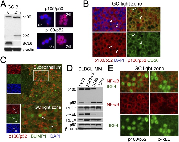
Expression and activation of alternative NF-κB subunits in normal and transformed human GC B cells and PCs. (A) Human tonsillar GC B cells ex vivo or following 24 h of coculture on CD40L-expressing feeders were subjected to Western blot analysis for p100/p52 and BCL6 (Left) and IF analysis for p105/p50 and p100/p52 (Right, red) and DAPI (blue). (B) IF analysis of tonsil sections for p100/p52 and DAPI or CD20 in the GC LZ. (C) IF analysis of tonsil sections for p100/p52, BLIMP1, and DAPI in the subepithelium and GC LZ. (D) Western blot analysis of DLBCL and MM cell lines for p100/p52, RELB, c-REL, RELA, and p105/p50. (E) IF analysis of tonsil sections for IRF4 and NF-κB subunits (either p100/p52 or c-REL) in the GC LZ. (Magnification: A–C and E, 400×.)

