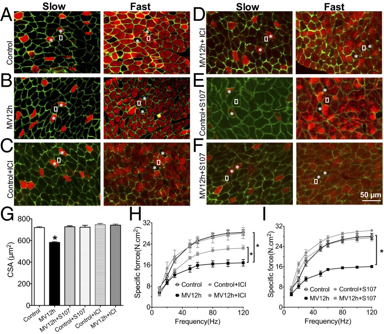
Fig. 6.
RyR1 dysfunction contributes to muscle fiber atrophy after 12 h of ventilation. Representative immunostaining of fast and slow diaphragm muscle fibers in mouse. Antibodies against fast- and slow-type myosin ATPase were used to perform immunostaining on cryosections of mouse diaphragm. Muscle membrane was counterstained with dystrophin antibodies. White squares indicate fast fibers, whereas the asterisks show the slow fibers in consecutive sections. Staining was performed in control (control) (A), mice under controlled mechanical ventilation during 12 h (MV12h) (B), control treated by ICI118551 (Control+ICI) (C), control mice treated by S107 (Control+S107) (D), mice ventilated during 12 h and treated by ICI118551 (MV12h+ICI) (E), and mice ventilated during 12 h and treated by S107 (MV12h+S107) (F). (G) Quantification of cross-section are in each condition (n = 192–852 fibers for each group, *P > 0.05). (H) Diaphragm muscle-specific force–frequency relationships recorded in control (n = 7), control+CI118551 (n = 3), MV12h (n = 10), and MV12h+ICI118551 (n = 6) (*P > 0.05, MV vs. control and MV12h+ICI). (I) Diaphragm muscle-specific force–frequency relationships recorded in control (n = 6), control+S107 (n = 3), MV12h (n = 6), and MV12h+S107 (n = 5) (*P > 0.05, MV vs. control and MV12h+S107).

