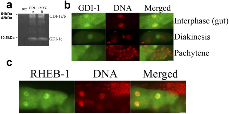
Fig. S5.
GDI-1 and RHEB-1 localize to both the nucleus and the cytoplasm. (A) Protein blot analysis of two lines expressing GDI-1 fused to a MYC tag. (B and C) animals expressing GDI-1::MYC (B) or RHEB-1::GFP (C) were stained with DAPI. Images show gonad (B) and gut (B and C) of young adult animals. (Scale bars: 10 µm.) Control WT animals stained with anti-MYC antibodies did not show specific staining.

