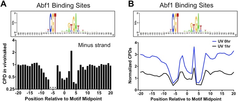
Fig. S6.
(A) Abf1-bound DNA sites show altered CPD formation. (Upper) The DNA consensus sequence of 661 Abf1 binding sites [generated using WebLogo (34)], including DNA flanking each binding site. (Lower) The scaled ratio of normalized CPDs in the UV 0-h sample (in vivo) relative to the UV 90J sample (naked) for the minus strands of Abf1 binding sites. Asterisks indicate that the indicated position in the motif cannot form CPD lesions because of DNA sequence constraints. (B) Comparison of normalized CPDs at Abf1 binding sites at 0-h and 1-h repair time points. The weighted average of normalized CPDs of both strands is depicted.

