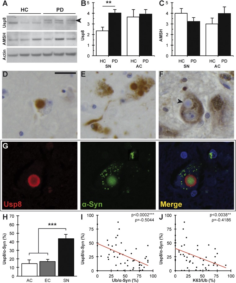
Fig. 2.
Usp8 expression and localization inversely correlate with ubiquitinated inclusions in α-synucleinopathies. (A) Representative immunoblot showing increased levels of Usp8 but not AMSH relative to the actin loading control in the SN from patients with LB disease; a specific band is indicated by an arrowhead. (B) Quantification of Usp8 protein level in the human brain showed a significant increase in the SN but not AC of patients with Lewy body disease compared with controls HC, healthy control (**P = 0.0028, n = 8). Quantification of AMSH protein levels did not show a significant difference (C). Usp8-positive LBs and Lewy neurites in the AC (D) and SN (E). (Scale bar, 20 µm.) (F) No AMSH staining was seen in nigral LBs, as indicated by the arrowhead. (G) Double immunofluorescence and confocal imaging confirmed the colocalization of Usp8 (red) and α-synuclein (green) in nigral LBs. DAPI indicates nuclear staining in blue. (H) Quantification of Usp8-positive as a percentage of α-synuclein–positive inclusions (Usp8/α-Syn) in serial sections showed a significant increase in the SN compared with cortical areas (AC, EC) (***P = 0.0001, n = 14). (I) Negative correlation between Usp8-positive and ubiquitin-positive inclusions in the SN (***P = 0.0002, ρ = −0.5044). (J) Negative correlation of Usp8-positive and K63-linked ubiquitinated inclusions shown as the ratio K63/Ub (**P = 0.0038, ρ = −0.4186). Error bars correspond to standard error of the mean.

