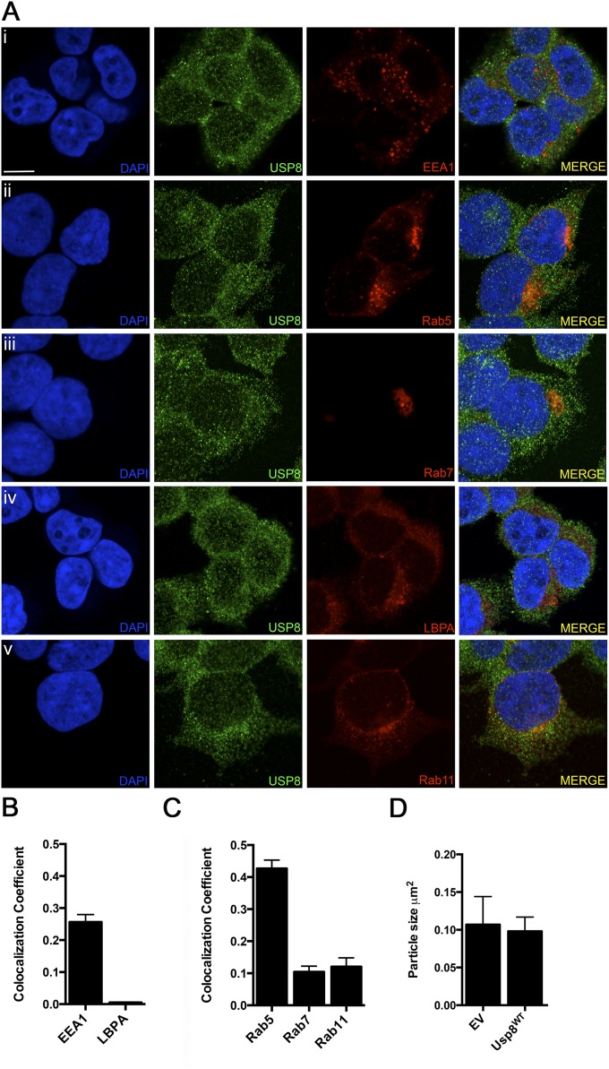
Fig. S4.
(A) Colocalization of Usp8 with various endosomal markers in HEK-293T cells. Cells were either transfected with fluorescent reporters or stained with specific antibodies: (i) early endosome, EEA1; (ii) early endosome, Rab5; (iii) late endosome, Rab7; (iv) late endosome, LBPA; and (v) recycling endosome, Rab11, and also costained for Usp8 (second column, green). (Scale bar, 10 μm.) (B) Quantification of the percentage overlap (MCC) of Usp8 with EEA1 and LBPA (endogenously expressed and stained using antibodies). (C) Quantification of the percentage overlap (MCC) of Usp8 with Rab5, Rab7, and Rab11 (fluorescent reporters transiently expressed). (D) Quantification of the size of EEA1-positive puncta corresponding to early endosomes in HEK-293T cells expressing FLAG-tagged wild-type Usp8 or an empty vector. Early endosome size was quantified by confocal microscopy [LSM 710 (Carl Zeiss)] and ImageJ, with n = 20 cells imaged per condition. The presence of untagged GFP within the vector enabled the identification of transfected cells. Error bars correspond to standard error of the mean.

