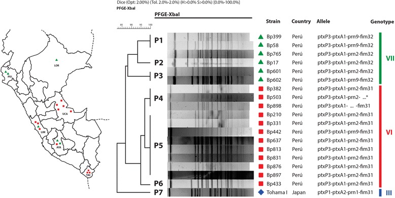
Fig. 3.

PFGE profiles and genotypes of B. pertussis Peruvian clinical isolates. Geographic distribution of B. pertussis isolates is shown on the left of the figure. The different individual patterns of pulse-field–gel-electrophoresis profiles in the center are indicated as P1 through P7. Genotyping was performed by sequencing the pertussis-toxin promoter (ptxP), pertussis-toxin subunit A (ptxA), pertactin (prn), fimbriae 2 and fimbriae 3. The classification of the allelic genotypes (indicated as Bp-strain numbers to the left of the sequences along with the country of origin)—III (blue), VI (red group), and VII (green group)—is based on the van Gent et al. (2012) report [44]
