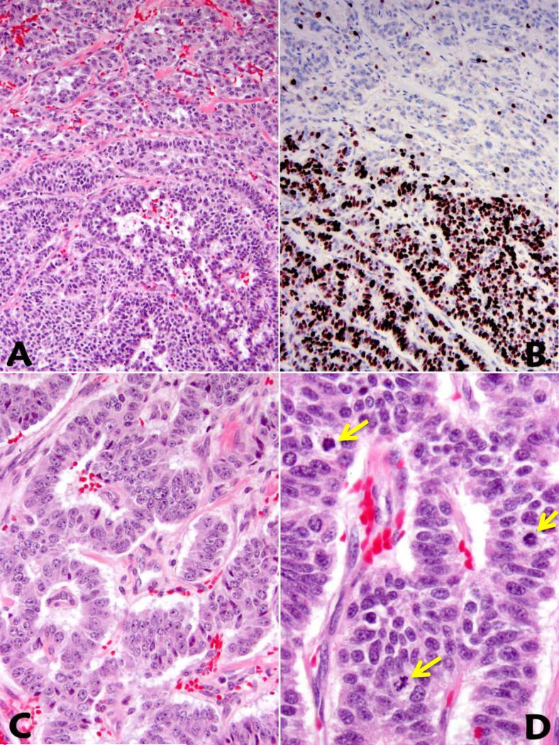
Figure 1.
Well-differentiated neuroendocrine tumor with HG component is characterized (in the direction from upper to lower) by subtle architectural alterations (A) and a markedly increased Ki67 proliferative index (B). In comparison with the lower grade component (C), areas with HG component within the same tumor reveal increased nuclear to cytoplasmic ratio and brisk mitotic activity (D).

