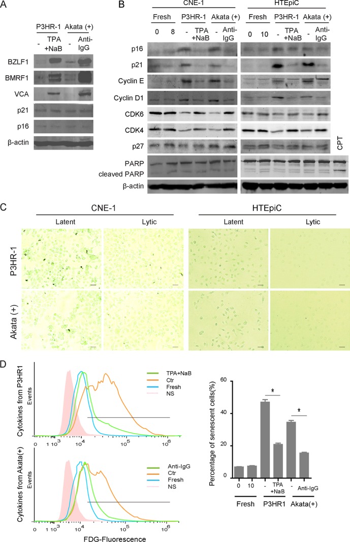
FIG 1.
Different patterns of paracrine senescence were observed during latent and lytic EBV infections. (A) EBV-positive P3HR-1 or Akata(+) cells were left uninduced or induced with 20 ng/ml TPA plus 0.3 mM NaB or 0.8% (vol/vol) anti-IgG for 24 h, respectively. The cells were collected, lysed, and analyzed by Western blotting as indicated. (B) Twenty-four hours later, the cells were washed twice and incubated with fresh medium for additional 24 h, and the cytokines secreted from uninduced (latent) or induced (lytic) P3HR-1 or Akata(+) cells were collected and used to generate conditioned media for NPC epithelial cells (CNE1) and primary epithelial cells (HTEpiCs), as indicated. After 8 or 10 days of culture with conditioned media, CNE1 and HTEpiCs were collected and analyzed using Western blotting. CPT, camptothecin. (C) The CNE1 cells and HTEpiCs were fixed, and senescent cells were detected with senescent β-Gal staining. Representative images under 200-fold magnification are shown, and the size bars represent 100 μm under microscopy. (D) After 10 days of culture with conditioned media, the HTEpiCs were digested, stained using an FDG probe, and measured using fluorescence flow cytometry. Representative images are shown (left), and the percentage of senescent cells was calculated from three independent experiments (right). NS, no-FDG-staining control. *, P < 0.01.

