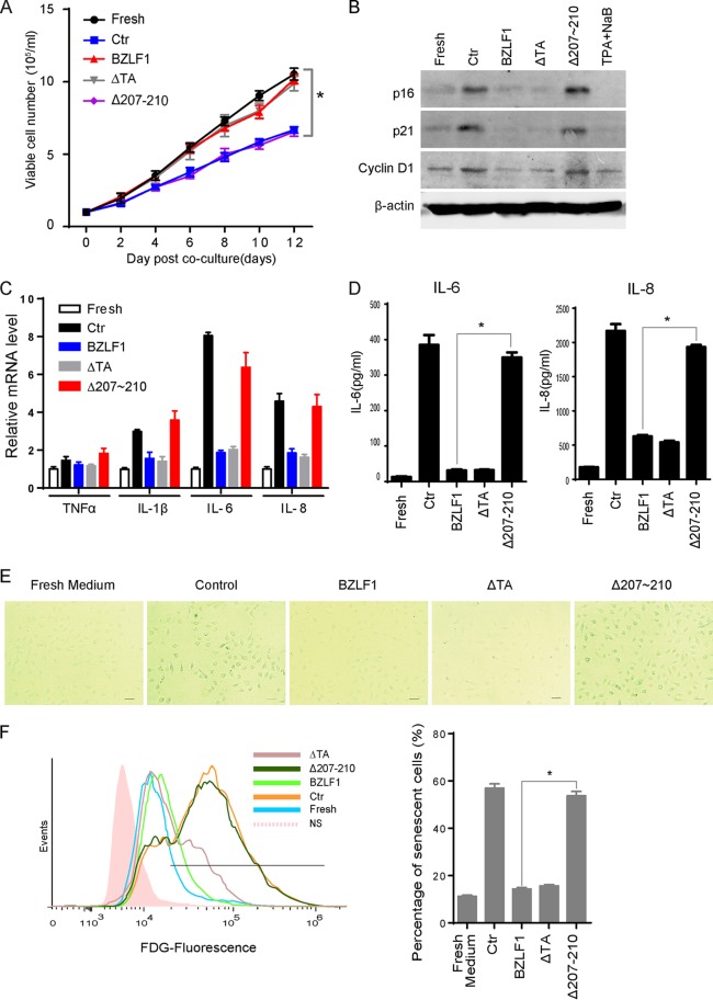
FIG 3.
BZLF1 blocked paracrine-mediated senescence and SASP. (A) Different cell growth was mediated by cytokines from P3HR-1 cells expressing BZLF1 or different mutants. HTEpiCs were seeded at low density (1 × 10⁴/ml) and then cultured with conditioned medium from control P3HR-1 cells or P3HR-1 cells overexpressing BZLF1, BZLF1ΔTA, or BZLF1Δ207-210. The conditioned medium was replaced every 2 days, and the living cells were digested and counted after trypan blue staining at different time points as indicated; the growth curves were recorded as the means of 5 random fields from three independent samples. Ctr, control. *, P < 0.01. (B) The cells were collected at 10 days after conditioned medium culture. p16INK4a, p21CIP1, and cyclin D1 expression levels were examined by Western blotting. (C) After 3 days of culture with conditioned medium, the cells were collected, and total RNA was extracted and analyzed by real-time PCR to detect expression of TNF-α, IL-1β, IL-6, and IL-8. (D) These cells were incubated with fresh FBS-free medium for 4 h, and then the titers of IL-6 and IL-8 in the medium were measured using ELISA kits. (E) After conditioned medium culture for 10 days, the cells were fixed, and β-Gal staining was used to detect activity of senescence-associated β-galactosidase. Representative images (200-fold magnification) are shown; the size bars represent 100 μm under light microscopy. (F) The senescent cells were determined using flow cytometry with FDG fluorescent staining, and the percentage of senescent cells was calculated from three independent experiments. *, P < 0.01.

