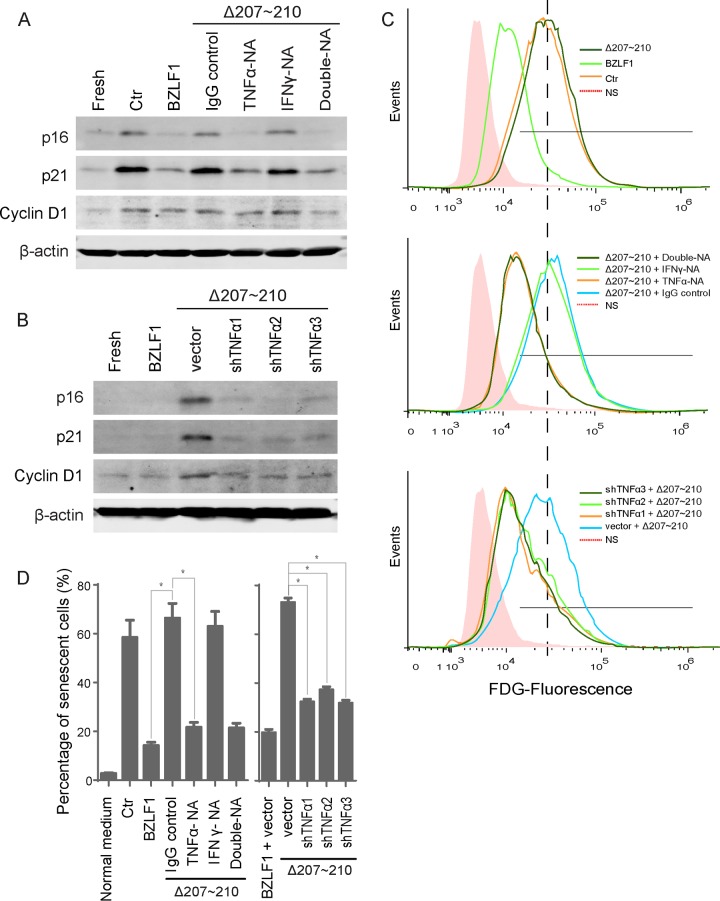
FIG 6.
TNF-α depletion ablated paracrine senescence. (A) Conditioned medium from P3HR-1 cells was neutralized with the anti-TNF-α or anti-IFN-γ antibody or control IgG before the medium was added to HTEpiCs. (B) Conditioned medium from P3HR-1 cells expressing TNF-α shRNAs was added to HTEpiCs, and the medium containing neutralizing antibodies was refreshed every 2 days. (A and B) The expression of cyclin D1, p21CIP1, and p16INK4a was examined after 10 days. (C) Representative images of fluorescent FDG-flow cytometry are shown. (D) The percentage of senescent cells from three independent experiments is shown. *, P < 0.01. NA, neutralizing antibody.

