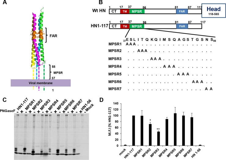
FIG 1.
Expression of MPSR triple alanine mutants. The 3× Ala mutants were made by mutations at the first 20 residues of the ectodomain of PIV5 headless HN1-117 to determine its functional significance. (A) Atomic model of PIV5 headless HN1-117 showing unresolved membrane-proximal stalk region (MPSR). F-interacting region (FAR) at the upper portion of the HN stalk is indicated. (B) Schematic diagram illustrating the PIV5 HN protein and showing the different domains: F-interacting region (blue), membrane-proximal stalk region (green), and transmembrane (TM) domain (red). The scheme of the 3× Ala mutants is shown. (C) Protein expression was determined using 30 min of 35S labeling followed by immunoprecipitation. Glycosylated species were digested by peptide-N-glycosidase F ([PNGase F]) treatment. Polypeptides were analyzed by 17.5% SDS-PAGE under reducing conditions. (D) Cell surface expression of HN1-117 and 3× Ala mutants was determined by flow cytometry. M.F.I., mean fluorescent intensity. Values are expressed as a percentage of HN1-117. Error bars represent standard deviations from three experiments. P values were calculated using Student's t test. *, P < 0.05; **, P < 0.01; otherwise, P > 0.05.

