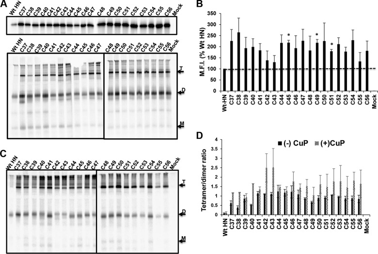
FIG 7.
The stalk of full-length HN can be trapped as disulfide-linked tetramers at the MPSR. (A) 35S metabolically labeled Cys mutants of full-length HN were resolved on a 10% SDS-PAGE under reducing (top) or nonreducing (bottom) conditions. Arrows indicate monomers (M), dimers (D), and tetramers (T) were 35S metabolically labeled. HN was immunoprecipitated, and polypeptides were analyzed by 10% SDS-PAGE. (B) Cell surface expression was quantified by flow cytometry and expressed as percentage of WT mean fluorescent intensity (M.F.I.). The horizontal dashed line represents the WT HN expression level. (C) WT HN and MPSR Cys mutants treated with CuP were resolved by nonreducing 10% SDS-PAGE. (D) Intensity bands of CuP-labeled proteins (C37 to C56) were quantified using ImageJ software, and the ratio of tetramer to dimer was estimated. *, P < 0.05; otherwise, P > 0.05.

