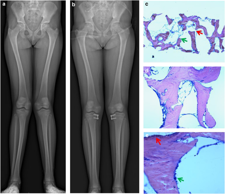
Figure 1.
Bone data in the first patient. (a) Radiographs of the legs at the time of bone biopsy: there is no evidence for abnormal mineralization; the bones are slightly bowed. (b) Radiographs of the legs after surgery: there is a complete correction of the deformations. (c) Bone biopsy at the tibia, May–Grünwald–Giemsa × 2.5: under low power, slightly impaired trabecular structure with focal trabecular separations (red arrow) and loose trabecular endings (green arrow) were seen. (d) Bone biopsy at the tibia, May–Grünwald–Giemsa × 10: mineralization appears normal. (e) Bone biopsy at the tibia, May–Grünwald–Giemsa × 20: mineralization appears normal, with active areas for both bone resorption (multinucleated osteoclasts) within resorption lacunae (green arrow) and bone formation (osteoblasts; red arrow).

