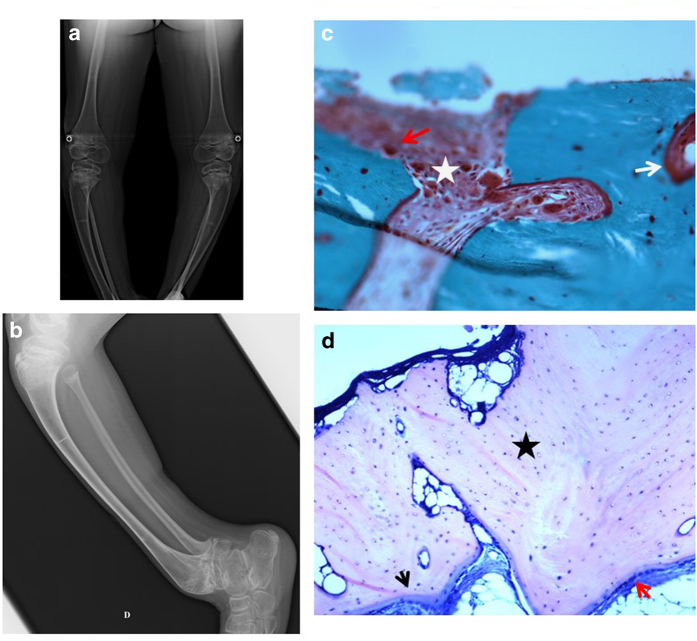
Figure 2.
Bone data in the second patient. (a) Radiographs of the legs 2 years before bone biopsy: severe deformations and sequelae of rickets. (b) Radiographs of the legs at the time of bone biopsy: severe deformations, sequelae of rickets and fractures. (c) Bone biopsy at the tibia, Goldner's trichrome, × 10: bone tissue with a partly fibrotic bone marrow (white star) and unusual resorption areas, with many multinucleated osteoclasts (red arrow). Mineralization appears normal (even though some part of the trabecular surface shows non mineralized osteoid, therefore leading to discuss a moderate surface osteoidosis; white arrow). (d) Bone biopsy at the tibia, May–Grünwald–Giemsa, × 10: elongated thin osteoid seams are colored in blue (black arrow) but mineralization appears normal. Numerous osteocytes (blue dots; marked with black star) are embedded in the mineralized matrix (pink), and some empty osteocytic lacuna (white dots) are observed.

