Figure 3.
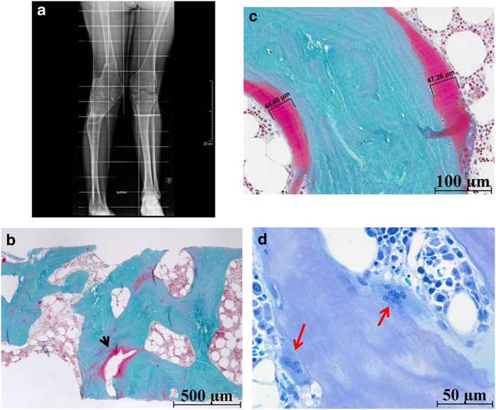
Bone data in the third patient. (a) Radiographs of the legs 8 months after bone biopsy: severe deformations, but no evidence of abnormal mineralization. (b) Goldner's trichrome, original magnification × 20: bone trabeculae showed focal deep defective mineralized matrix (red-stained bone regions; black arrow). (c) Goldner's trichrome, original magnification × 20: up to 50-μm-thick osteoid seams (red-stained bone regions) are observed on the surface of mineralized (green-stained portions of bone matrix) bone trabeculae. (d) Toluidine blue, original magnification × 40: enlarged osteoclasts contained up to eight isomorphic nuclei (red arrows). No marrow fibrosis was apparent in this specimen.

