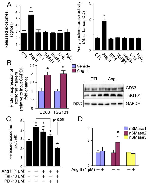
Fig. 3.
Ang II-induced release of exosomes from cardiac fibroblasts. (A, B) The effect of pathological stress on the release of exosomes from cardiac fibroblasts. (A) The amount of proteins and the activity of acetylcholinesterase in total released exosomes from the same number of neonatal rat cardiac fibroblasts that were stimulated with vehicle, Ang II (1 μM), ET-1 (0.1 μM), TGFβ1 (10 ng/ml), insulin (0.1 μM), LPS (0.1 μg/ml), H2O2 (500 μM) as indicated for 48 h. n=3, *p<0.05 vs. vehicle-treated control (CTL). (B) Western blot analysis of CD63 and TSG101 in total exosomes from the same number of neonatal rat cardiac fibroblasts that were stimulated with vehicle or Ang II for 48 h. Left panel shows semi-quantified results. n=3, *p<0.05 vs. vehicle-treated control (CTL). Right panel is the immunoblots (n=3). Input: 10 μl of the neonatal rat cardiac fibroblast lysates. (C) The effect of Telmisartan and PD123319 on Ang II-induced secretion of exosomes from cardiac fibroblasts. Neonatal rat cardiac fibroblasts were treated with Ang II (1 μM), Telmisartan (Tel, 10 μM), PD123319 (PD, 10 μM) as indicated for 48 h. The total proteins of exosomes secreted were measured by BCA assay and normalized by cell number. n=3, *p<0.05 vs. vehicle treated control (−). (D) The effect of Ang II on mRNA expression of neutral sphingomyelinase (nSMase) in cardiac fibroblasts. Neonatal rat cardiac fibroblasts were treated with vehicle of Ang II (1 M) for 48 h and subjected to qPCR analysis of nSMase1, 2, and 3 expression. n=4, *p<0.05 vs. vehicle-treated control (−).

