
Figure 3.
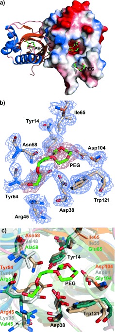
Crystal structure of SalBIII. a) Overall fold of SalBIII. The right‐hand monomer is shown with its electrostatic surface (blue: negatively charged; red: positively charged). PEG: polyethylene glycol. b) Predicted active site residues in SalBIII. c) Superposition of the active sites of SalBIII (orange), Cyc11 (model, gray) and Lsd19 (green).