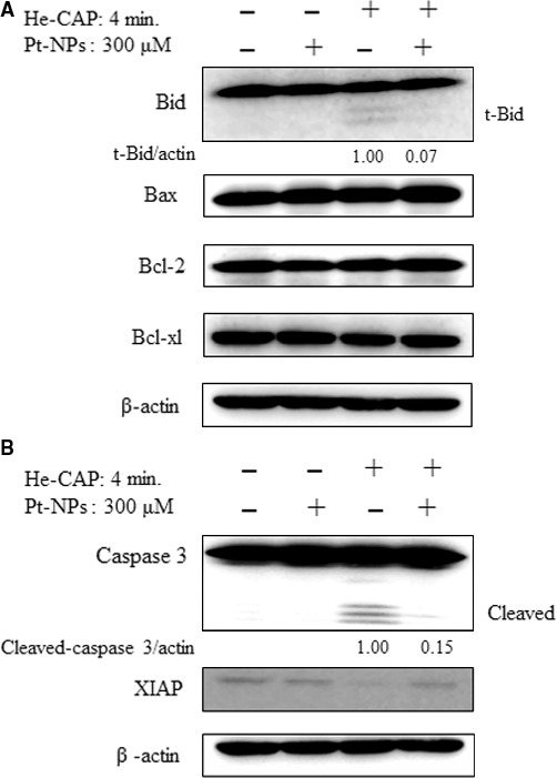
Figure 6.

Assessment of apoptosis‐related proteins. Cells were treated with or without Pt‐NPs and, then harvested 18 hrs after helium‐based cold atmospheric plasma treatment. (A) Western blot analysis of Bcl‐2 family proteins (B) Changes in the expression levels of caspase‐3 and Xiap as detected by Western blot analysis. The t‐Bid and cleaved caspase‐3 signals were normalized to β‐actin and the relative ratios are shown below the band. Band density was evaluate by Image J software and expressed as fold change.
