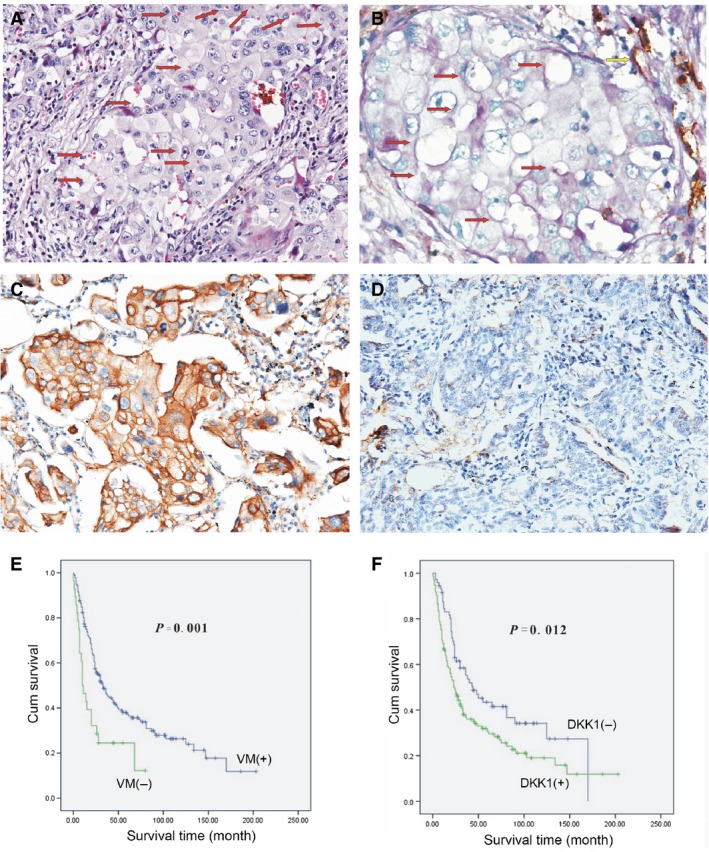
Figure 1.

(A) Morphological appearance of VM with hematoxylin and eosin staining. The VM channel was surrounded by tumour cells and RBC (red arrow). Absence of necrosis and phlogocyte was observed in the vicinity (×200). (B) Results of CD31/PAS double‐staining (×400). The VM channel (red arrow) was PAS‐positive, but it did not expressed CD31. Endothelium‐dependent vessel (yellow arrow) was PAS and CD31‐positive. (C) Immunohistochemical staining for DKK1 overexpression in the VM‐positive group (×200). (D) DKK1 was down‐regulated in the non‐VM group (×200). (E) Kaplan–Meier survival analysis showing that the VM‐positive patients have shorter survival time than VM‐negative patients (P = 0.001). (F) Kaplan–Meier survival analysis showing that the DKK1‐positive patients have shorter survival time than DKK1‐negative patients (P = 0.012).
