
Figure 5.
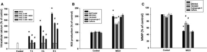
Reciprocal amplification of ROS production and intracellular calcium increase in MGO‐treated ARPE‐19 cells. (A) Cells were pre‐incubated with NAC (2 or 5 mM) for 30 min and then incubated with 300 μg/ml MGO for 1, 3 or 6 h. Intracellular calcium levels were determined by using Fluo‐3 AM and flow cytometry. (B, C) Cells were pre‐treated with BAPTA/AM (10 μM), MRS1845 (10 μM), YM‐58483 (10 μM), xestospongin C (1 μM) or caffeine (10 mM) for 30 min and then incubated with 300 μg/ml MGO. After 1 h, cellular ROS levels were measured with H2 DCFDA by using flow cytometry (B). After 3 h, MMP was measured with rhodamine 123 by using flow cytometry (C). *P < 0.05, indicating the significant effects of MGO. # P < 0.05, indicating the significant inhibition of MGO responses by indicated agents.