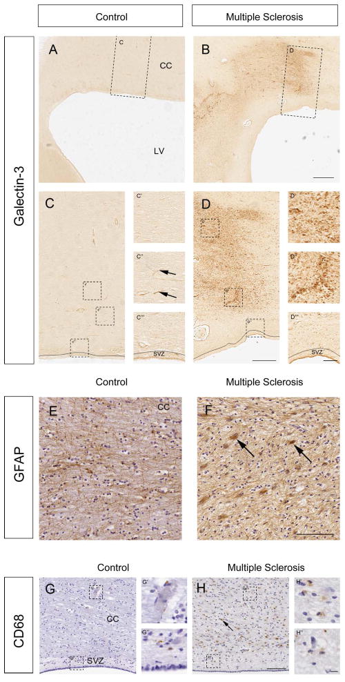
Fig. 1. Periventricular regions express Gal-3+ in human MS.
(A,C) Gal-3 immunoreactivity was low surrounding the lateral ventricles (LV), and in the corpus callosum (CC) of controls. Boxed areas shown at higher magnification in C-C‴. Arrows show blood vessels. SVZ outlined in C and C‴. (B) Gal-3 expression was markedly increased in periventricular regions in MS cases. Boxed area shown at higher magnification in D-D‴. (E) GFAP immunoreactivity in the corpus callosum of a control human. (F) GFAP+ reactive astrocytes (ex. arrows) in the corpus callosum of an MS case. (G) CD68 immunoreactivity in a control human section. The SVZ is well delineated in this section by the lower cell density seen with haematoxylin counterstain in the hypocellular gap. Boxed areas shown in G′ and G″. (H) Many CD68+ cells (ex. arrow) were detected in the corpus callosum of a human with MS. Boxed areas shown in H′ and H″. All panels are of sections taken in the coronal plane. Scale bars: B=1 mm; D=500 μm; D‴=100 μm; F, H=100 μm; H″=10 μm.

