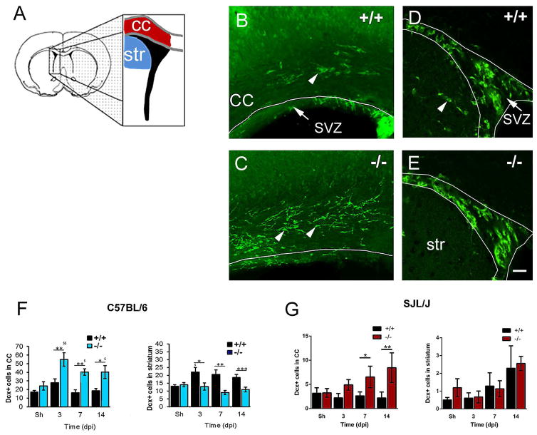
Fig. 6. The number of Dcx+ cells in the corpus callosum was greater in Gal-3−/− mice after TMEV infection.
(A) Forebrain schematic showing location of analysis for SVZ neuroblast emigration into the corpus callosum (cc) and striatum (str). (B–C) Dcx immunofluorescence in the corpus callosum and SVZ of C57BL/6 WT and knockout mice. Arrowheads point to Dcx+ cells in the corpus callosum. (D–E) fewer Dcx+ cells were found in the striatum of C57BL/6 Gal-3−/− mice after TMEV. Arrowhead points to Dcx+ cell in the striatum. Scale bar: F=20 μm. (F) The number of Dcx+ cells increased in the corpus callosum in C57BL/6 Gal-3−/− mice compared to WT. The number of Dcx+ cells decreased in the striatum in C57BL/6 Gal-3−/− mice compared to WT. (G) The number of Dcx+ cells increased in the corpus callosum in SJL/J Gal-3−/− mice compared to WT SJL/J. The number of Dcx+ cells in the striatum in SJL/J Gal-3−/− mice was not significantly different compared to WT SJL/J’s.*P<0.05 Gal-3−/− compared to WT, ** P<0.01, ***P<0.001, $ P<0.05 TMEV treated compared to shams, $$ P<0.01.

