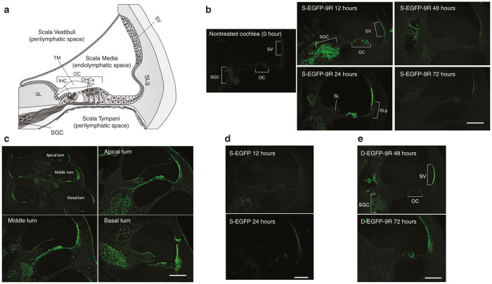
Figure 1.
Sectional images after enhanced green fluorescent protein (EGFP) or EGFP-9R transduction. (a) Cross-sectional diagram of a normal adult cochlea. The adult mammalian cochlea consists of three compartments: the scala vestibuli; scala tympani; and, scala media. The scala media contains the organ of Corti (OC). The OC contains inner hair cells (IHCs) and outer hair cells (OHCs). The IHCs and outer hair cells (OHCs) act as mechano-electrical transducers and play a crucial role in hearing. The electrical signal is transmitted via the spiral ganglion cells (SGC) to the auditory pathway of the brain. The stria vascularis (SV), located in the lateral wall of the scala media, is responsible for the secretion of K+ into the endolymph and the production of the endocochlear potential. (b) The images show sections of the middle turns of the cochleae. In the s-EGFP-9R group, EGFP was detectable strongly in the SV, OC and SGC at 12 hours. Also EGFP was detectable strongly in the SV, OC, and SGC, and slightly in the spiral limbus (SL) and the spiral ligament (SLg) 24 hours. EGFP levels were moderately detectable at the SV and SG at 48 hours and only slightly detectable at the SV at 72 hours. (c) At 24 hours in the s-EGFP-9R Group. EGFP was detectable at all turns in the cochlea. (d) At the middle turn of the cochleae in the s-EGFP group, EGFP was slightly detectable in the SGC at 12 hours and in the SV at 24 hours. (e) At the middle turn of the cochleae in the d-EGFP-9R group, EGFP was significantly detectable in the SV and SGC at 48 hours. The scale bars indicate 50 μm.

