Figure 5.

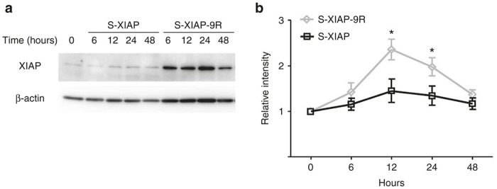
Chronologicanl changes of XIAP protein expression levels. (a) Representative western blot results showing the transduction levels of X-linked inhibitor of apoptosis protein (XIAP) in the whole cochleae. (b) Quantification of the transduction levels of XIAP in the cochleae. The transduction levels for XIAP in s-XIAP-9R-treated cochleae were significantly higher than those in XIAP-treated cochleae at 12 and 24 hours. Each n = 4. *P < 0.05.
