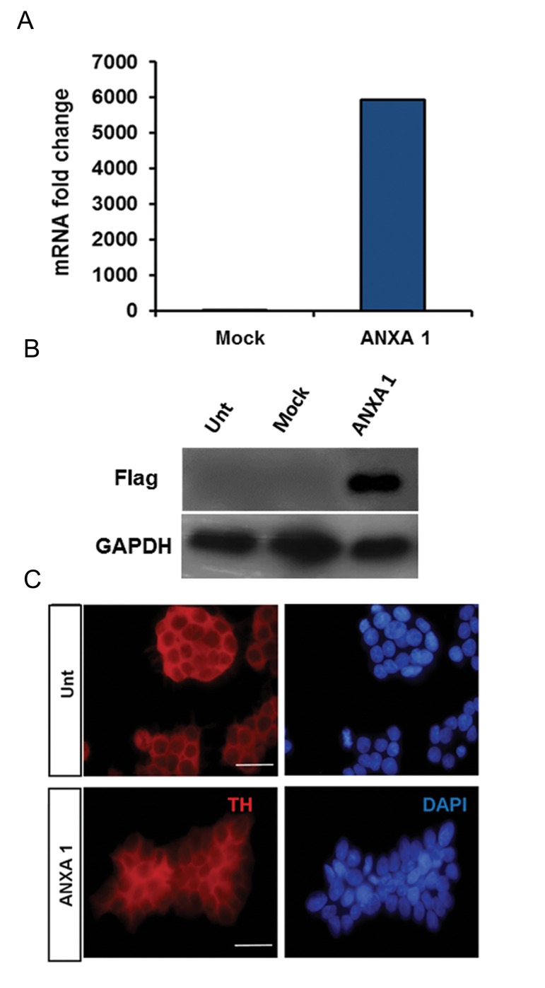
Fig.1.
Characterization of ANXA1-FLAG transfected PC12 cells. A. Ectopic expression of ANXA1-FLAG. Relative expression of ANXA1-FLAG was quantified and normalized to the amount of GAPDH transcript, B. Western blot analysis to quantify the protein content of ANXA1-FLAG. GAPDH was used as a loading control, and C. PC12 cells were stained with an antibody against TH. Nuclei were counterstained with 4′, 6-diamidino-2-phenylindole (DAPI). Bar is 200 µm. It should be noted that the staining pattern in ANXA1 transfected cells was similar to untransfected cells (Unt). ANXA1 and Mock represent stably transfected cells with pEPi FGM18F PGL-268/ANXA1-FLAG and pEPi FGM18F PGL268 plasmids respectively. ANXA1; Annexin A1.

