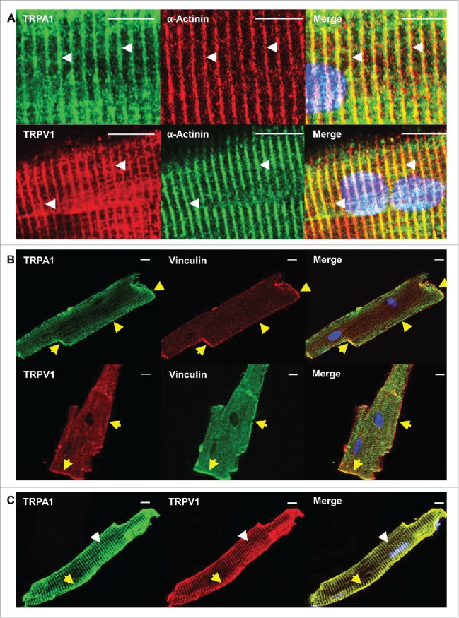
Figure 4.

Confocal images in CM obtained from WT mice confirm that TRPA1 and TRPV1 colocalize at the Z-disc, costameres as well as the intercalated discs. Freshly isolated CMs were double labeled with antibody recognizing TRPA1 or TRPV1 and α-actinin (z-disc marker; white arrows) A). Similar immunolabeling with antibody recognizing TRPA1 or TRPV1 and vinculin (costamere and intercalated disc marker, yellow arrow) was also performed (B). Representative confocal images of double-labeled CMs revealed that TRPA1 and TRPV1 colocalize at the Z-disc, costamere, and intercalated discs (C). Scale bar, 10 μm.
