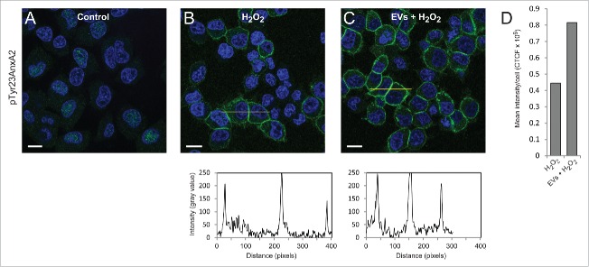
Figure 2.
Pre-incubation of PC12 cells with EVs derived from H2O2-treated cells results in ∼2-fold higher levels of pTyr23AnxA2 upon subsequent treatment with H2O2, as compared to cells exposed to H2O2 only. PC12 cells were untreated (A), treated for 15 min with 1 mM H2O2 only (B), or treated for 15 min with 1 mM H2O2 after their pre-incubation for 2 h with EVs released from cells exposed for 1 h to H2O2 (C). pTyr23AnxA2 (green, A-C) was detected using monoclonal antibodies. The DAPI-stained nuclei are shown in blue. Scale bars: 10 μm. The diagrams below each panel show the corresponding intensity profiles along the lines indicated in the respective panels. Panel 2D: Corrected total cell fluorescence (CTCF) was measured from cells (20–30) in the samples shown (panels A–C); CTCF were then divided by the number of cells measured.

