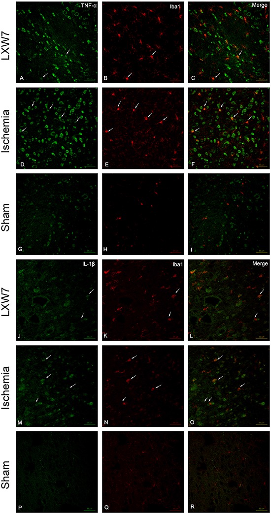
Figure 4. Treatment of middle cerebral artery occlusion rats with LXW7 (LXW7 group) resulted in the reduction of TNF-α, and IL-1β expression in activated microglia. Confocal images showing the expression of TNF-α (A, D, G, green), IL-1β (J, M, P, green) in microglia (B, E, H, K, N, Q, red), in peri-ischemic brain tissue 24 h after MCAO in rats (D-F, M-O) and following treatment with LXW7 (A-C, J-L) (n=6 for each group). Expression of TNF-α and IL-1β in microglia cells (arrows) is markedly enhanced following MCAO, but a noticeable reduction in TNF-α and IL-1β expression can be observed in the activated microglia in the LXW7-treated rat brain. Activated microglia were reduced with LXW7 treatment (B, K). Scale bars: 50 µm.

