Fig. 3.
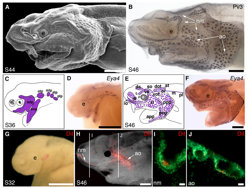
Lateral line placodes give rise to ampullary organs and neuromasts in a basal ray-finned bony fish, the North American (Mississippi) paddlefish, Polyodon spathula. Lateral views, anterior to the left, unless otherwise noted; staging according to Bemis and Grande (1992). All panels were previously published in Modrell et al. (2011a) and are reproduced here in accordance with the terms of the authors’ Licence to Publish agreement with Nature Publishing Group. (A) Scanning electron micrograph of a stage 44 embryo showing differentiated ampullary organ fields, particularly on the operculum. (B) Stage 46 embryo immunostained for the Ca2+-binding protein parvalbumin-3 (Pv3), which is strongly expressed in the sensory receptor cells of both neuromasts and ampullary organs (also see Modrell et al., 2011a). (C-F) Schematic diagrams and whole-mount in situ hybridisation for the transcription co-factor gene Eya4 at (C,D) stage 36, when Eya4 is expressed in developing neuromast canal lines and the ampullary organ fields flanking those lines (purple in C) and (E,F) stage 46, when Eya4 expression is maintained in both neuromasts and ampullary organs (purple in E). (G) Stage 32 embryo immediately following a focal DiI injection into the anterodorsal lateral line placode (injection site outlined in red). (H) The same embryo as in G, at stage 46. DiI-labelled cells are visible both in a neuromast canal line and ampullary organ fields. Lines indicate the plane of transverse sections showing DiI-labelled cells (red) in (I) a neuromast and (J) ampullary organs, both counterstained with the nuclear marker Sytox Green (green). Abbreviations: adp, anterodorsal placode; ao, ampullary organ; app, anterior preopercular ampullary field; avp, anteroventral placode; dot, dorsal otic ampullary field; di, dorsal infraorbital ampullary field; ds, dorsal supraorbital ampullary field; e, eye; epi, epibranchial placode region; io, infraorbital lateral line; m, middle lateral line; mlp, middle lateral line placode; ol; otic lateral line; otp, otic lateral line placode; plp, posterior lateral line placode; pll, posterior lateral line; pop, preopercular lateral line; ppp, posterior preopercular field; S, stage; stp, supratemporal placode; so, supraorbital lateral line; st, supratemporal lateral line; vi, ventral infraorbital field; vot, ventral otic field; vs, ventral supraorbital field. Scale bars: (A,B,D,G) 0.5mm, (F,H) 1mm, (I,J) 10μm.

