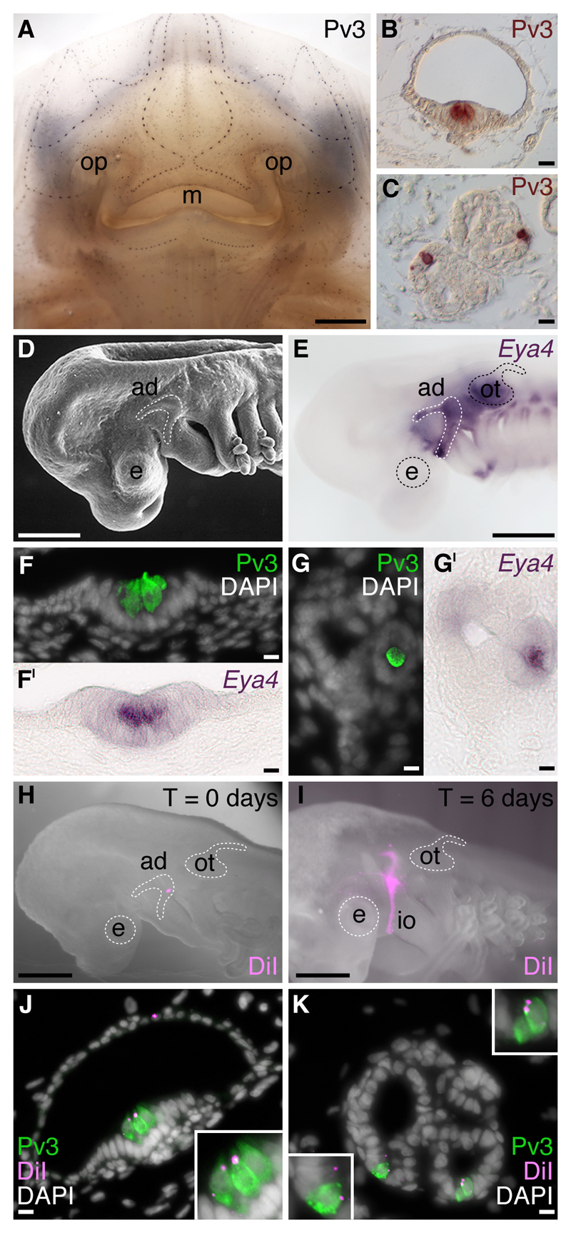
Fig. 4.
Lateral line placodes give rise to ampullary organs and neuromasts in a cartilaginous fish, the little skate, Leucoraja erinacea. All panels except E, H and I were previously published in Gillis et al. (2012) and are reproduced here in accordance with the terms of the authors’ Licence Agreement with the Company of Biologists. (A) Whole-mount immunostaining for the Ca2+-binding protein parvalbumin-3 (Pv3) in an L. erinacea embryo at stage 33 (Maxwell et al., 2008) reveals superficial lines of cephalic mechanosensory neuromasts, as well as clusters of ampullary organs located deeper within the dermis. Immunohistochemical localisation of Pv3 in (B) neuromasts and (C) ampullary organs reveals small clusters of Pv3-positive sensory receptor cells nested among Pv3-negative supporting cells. To test the hypothesis that lateral line placodes give rise to neuromasts and ampullary organs, we fate-mapped the anterodorsal lateral line placode in L. erinacea, which is recognisable (D) as a horseshoe-shaped thickening of cranial ectoderm caudal to the eye and dorsal to the mandibular arch, and (E) by its expression of the transcription co-factor gene Eya4. Eya4 expression is maintained at later stages in the Pv3-positive sensory receptor cells of (F,FI) neuromasts and (G,GI) ampullary organs. (H) Example of an embryo immediately after focal labelling of the anterodorsal lateral line placode with the lipophilic vital dye DiI. (I) After 6 days of incubation, DiI-positive cells were observed migrating away from the placode, in the infraorbital sensory primordium. In embryos with DiI-labelled anterodorsal lateral line placodes, sensory receptor cells, support cells and canal cells of (J) neuromasts and (K) ampullary organs were DiI-positive, indicating their lateral line placodal origin. Abbreviations: ad, anterodorsal lateral line placode; ad, anterodorsal lateral line placode; e, eye; io, infraorbital sensory primordium; m, mouth; op, olfactory pit; ot, otic vesicle. Scale bars: (A) 2.5mm, (B-C) 10μm, (D,E,H) 0.5mm, (I) 0.4mm, (F-G’,J,K) 10μm.

